Advantages of performing a pre-extraction assessment of hopeless teeth prior to treatment planning extractions and dental implants.
Inadequate bone in potential dental implant sites can present with major concerns for dentists that place or restore dental implants because often implants placed in inadequate bone can result in angulation problems to compensate for inadequacy, progressive bone loss, or even an inability for implant to be placed or integrate. Therefore, it is essential that if planning to place dental implants a thorough assessment is performed. This usually begins in the treatment planning phase prior to tooth extraction.
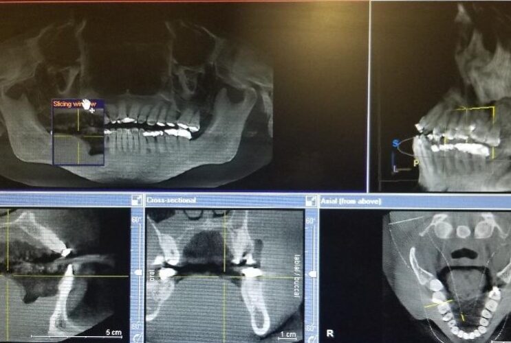
Assessment of bone around hopeless teeth and edentulous sites involve an evaluation in 3 dimensions, looking at bone thickness, width and height assessed in the facio-lingual, mesiodistal, and apico-coronal dimensions. The goal is to schedule procedures that would correct bone deficiencies when hopeless teeth are being extracted so as to maximize chances for having optimal bone present post extraction.
Completing a pre-extraction clinical and radiographic assessment using 3D cone beam x-rays or panoramic x-rays combined with sounding for changes in topography, as well as evaluating articulated models of the site allow comprehensive planning. As a result, procedures such as socket preservation, ridge augmentation, soft tissue grafting or orthodontic extrusion can be performed to maximize bone and soft tissue in the future implant site. The assessment also allows determination of type of implant placement protocol to utilize.
Additionally, buccally located teeth tend to have thinner facial plates, with extraction sites prone to dehiscence defects and loss of buccal plate during extraction. As a result, performing a pre-extraction assessment would allow treatment planning of the necessary procedures that can be able to correct some of these deficiencies prior dental implant placement.