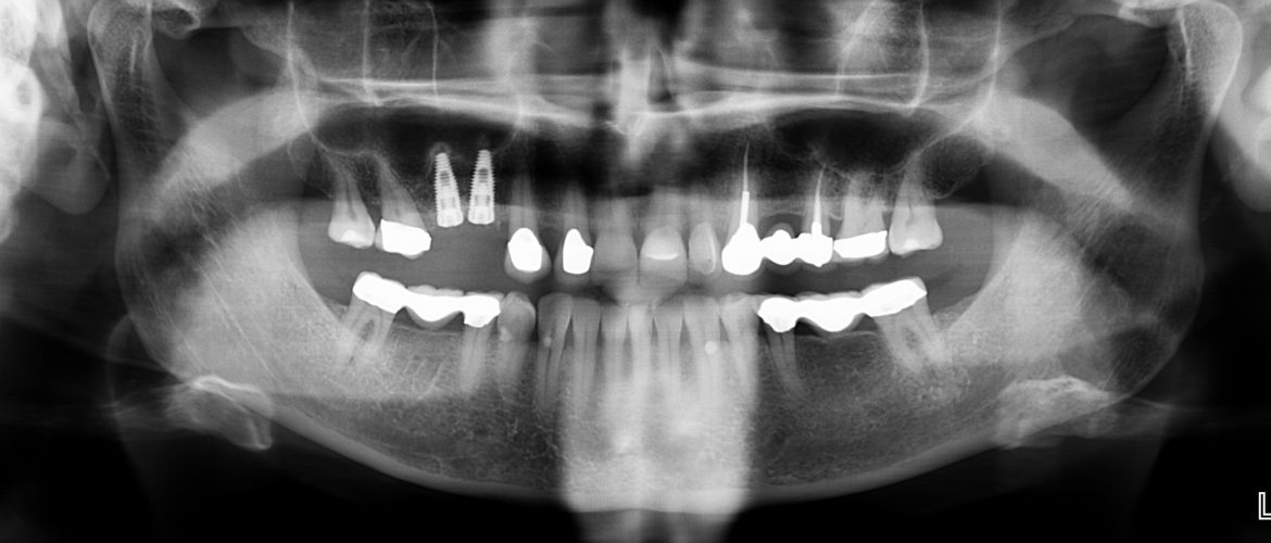
Osteotome Technique

Review Article on Osteotome technique: A minimally invasive way to increase bone for dental implant placement in the posterior maxilla and prevent sinus membrane perforation for single and multiple teeth replacements. Dr. Nkem Obiechina
Abstract:
The posterior maxilla presents with limitations due to the presence of the maxillary sinus as well as overall poor bone quality due to decreased bone density. As a result, procedures such as sinus and ridge augmentations are often required following tooth loss to prepare future implant sites for dental implant placement. The methods that are available for sinus augmentation involve the lateral (direct) approach and the crestal (indirect) approach. While a number of studies find comparable results for bone gain after sinus augmentation, the lateral approach presents with limitations including post op morbidity, and limited access for single tooth areas as well as higher potential of Schneiderian membrane damage. As a result, the crestal approach has been advocated. A number of modifications have been made to the approach to improve its efficacy since it was described by Summers in 1994 and the goal of this article is to review some of these improvements and how they have affected overall success of the technique.
Introduction:
Prolonged tooth loss can result in loss of bone in edentulous sites in the mouth especially in the posterior maxilla. Anatomical sites in the mouth that are close to areas with bone loss are at particular risk of being encroached on during dental implant placement. Vital structures such as the maxillary sinus, mental and inferior alveolar nerves have to be identified, and modifications such as use of shorter implants, and sinus augmentation for the posterior maxillary region have been implemented as ways of circumventing possible damage to these structures.
The posterior maxilla presents with medullary bone which has less density and quality than the premaxilla and the mandible.1 In addition, pneumatization that occurs in the sinus area after extraction due to osteoclastic resorption at the maxillary posterior ridge as well as increased positive pressure of the maxilla overtime, results in significant decrease of bone volume present, necessitating need for sinus lift and ridge augmentation in the posterior maxilla.2
 The initial approach to sinus augmentation involved the use of the lateral approach discovered by Tatum in 1977 and further published by Boyne in 1980.3 While the technique has provided the advantage of being able to augment sites with multiple implants simultaneously, a number of drawbacks exist such as increased post-operative morbidity such as swelling and hematoma formation4. Other limitations of the lateral sinus lift approach include increased need for visibility and access to the sinus, potential problems in sites with limited access, as well as potential of damage to the Schneiderian membrane.3 All of which necessitated the creation of crestal access to perform sinus augmentation due to reduced postoperative morbidity and less invasive technique.2,3 The Lateral window is usually the recommended technique for severely atrophic maxillary ridges due to its ability to be able to augment a wider area of bone at one time.1,2,3,4
The Osteotome technique was first detailed in multiple publications by Summers where use of blunt instruments called osteotomes were used for elevation of the sinus, bone augmentation occurs followed by dental implant placement simultaneously or four to six month later as a two stage technique.5 Modifications to the technique involves simultaneous sinus augmentation and implant placement which has been found to have a high survival rate comparable to dental implant placement in native bone.1,2 Other modifications involve use of modified osteotomes 17and also use of a hydraulic balloon for sinus augmentation.18,19,20
The osteotome technique is also used for site development and widening ostectomy sites with limited bone present. Overtime the technique has been modified and its use expanded including increasing density of bone by using the osteotomes to compact bone is areas with porous bone, and atraumatically widening ostectomy sites.3 It therefore presents with major advantages over the lateral window technique especially in single edentulous sites that are being replaced with dental implants.
Advantages of the osteotome technique include that the ostectomy site is much smaller so less post operative morbidity and less healing time needed, it is more efficacious, and has the ability to increase bone to implant contact by ability to compact bone increasing density, and therefore can result in more stability around implants.6 It is also able to allow access to areas of the mouth were the lateral window would have limited access such as single implant placement sites as well as areas of the mouth with restricted access or risk of membrane perforation. 6


A number of studies have suggested the crestal approach to sinus augmentation using osteotomes to replace the lateral window technique for single tooth implant replacement in the posterior maxillary where due to impeded access there is high possibility that perforation of the Schneiderian membrane can result due to the limited access, and there can be an increased risk of post op morbidity with the lateral window technique.3
Studies comparing the efficacy of the lateral window and osteotome technique found comparable results in terms of stability of implants that were placed, comparable new bone gain after grafting procedure ranging form 4-8mm from both techniques as well similar survival rates as found with native non augmented maxillary bone.1,2 While the lateral window technique resulted in more swelling than the osteotome technique, it was found to be more efficacious for severely atrophic maxilla with less than 3mm of bone present. 1,2 The osteotome technique was however found to be minimally invasive with higher patient acceptance.2
Use of the osteotome technique especially in the posterior maxilla also offers the benefit for improving direct contact between implant and bone because of the ability of osteotomes to compress bone with reduced bone density (type 3 and type 4 bone) allowing better anchorage of the dental implants by forming a denser connection between the compacted bone and implants.3,5 Types of osteotomes have also been modified with the creation of expansive osteotomes which are similar to the original Summers osteotomes except additional features such as apical tip design, different calipers that are designed to adapt to different systems, as well angulated osteotomes which have been designed to improve bone density and lateral compression of bone, and help improve primary implant retention and stability.7High success rates of 95% to 97% were reported with these osteotomes.7
Modifications to the Osteotome techniques:
The requirements for simultaneous implant placement with sinus augmentation versus delayed placement include that to reduce risk of damage to the Schneiderian membrane, implants could be placed at time as sinus elevation when a minimum of 5mm of bone is present and as a delayed protocol when less than 5mm of bone is present.1, 2
More recently some studies have reported successful use of osteotome technique for atrophic maxilla where usually the lateral window technique would have been the cause of action8 Anson et al found successful implant stability and high survival rates in sites with 2-3mm of native bone present with osteotome use combine with a composite graft comprised of demineralized freeze dried bone allograft, bovine bone and calcium phosphate.9
Toffler developed the crestal core elevation technique (CCE) for sinus augmentation using osteotomes which involves using a delayed approach to place multiple implants in atrophic posterior maxilla with less than 5mm of native bone present. The technique involves initial access using a trephine followed by osteotome site augmentation using #5 and #6 core osteotomes and then by augmentation of the sinus using a composite autogenous graft combined with bovine bone, followed by an e-PTFE membrane.10 The area is allowed to heal for about 5-7 months before dental implant placement occurs. Bone gain from technique ranges from 7-12mm was reported, it also offers a less invasive technique than the lateral window, and minimizes chances of Schneiderian membrane exposure.10
Narang et al evaluated the use of modified osteotome sinus floor elevation combined with platelet rich fibrin, bone grafts and immediate implant placement and found high survival results for immediate dental implants placed in the posterior maxilla using the osteotome technique.11
The Balloon sinus lift was also implemented for use in atrophic maxilla, and involves drilling of the ostectomy site following the use of osteotomes to 1mm from the Schneiderian membrane followed by insertion of a latex balloon attached to a catheter to lift the sinus floor with slow infusion of saline and then placement of bone grafting material for sinus augmentation with increased bone gain compared to the osteotome technique alone. 17, 19 Penaroccha-Diago et al described the technique and recommended its use in sites with 3mm or more native bone present. They combined the balloon lift with bovine bone mixed with autogenous bone along with simultaneous dental implant placement and indicated that they had a 100% success rate after 1 year of dental implant loading.19 They indicated its advantage of increasing bone after sinus lift by 8.7-10mm over the 3-4mm gain from the standard osteotome technique making its use invaluable in the atrophic maxilla.19,20
A number of studies have evaluated the effect of not using any bone grafting materials for osteotome augmentation and found comparable to results to use of bone grafting materials. 12,13 Taschieri et al found a success rate of 98.02% at 1 year and 96.77% after 5 years for 1767 implants evaluated without use of bone grafts for osteotome sinus augmentation.12 Brizuela et al found a success rate using graftless technique of 91.6% after 2 years of patient follow up.14
Similar results were found for use of a graft-less lateral window approach, Falah et al reported a 94% success rate for dental implants when fibrin blood clot was utilized instead of bone grafting material for sinus augmentation, their conclusion is that the blood clot serves as an osteoprogenitor which affects migration, differentiation of bone cells as well as regeneration of bone.15Pichasov and Juodzbalys evaluated a number of studies between 1993 and 2013, involving use of graft-less sinus lift technique and found that all the articles reviewed found increased bone formation and high implant stability and survival rate using the patients’ blood clot instead of bone grafts.16
Conclusion:
The osteotome technique continues to be an efficacious way to effectively achieve bone augmentation in the posterior maxilla with reduced post-operative morbidity and relatively quicker healing compared to the lateral window technique. Its limitation compared to the lateral window technique is usually with severely atrophic maxillas that present with less than 4mm of native bone as well as where multiple implants are being placed to replace teeth. A number of modifications have been made to the technique to overcome these limitations, with success noted, but further research is needed to be able to evaluate immediate placement with atrophic maxillas as well as use of graft-less technique with use of patient’s blood instead of bone grafts for osteotome sinus augmentation.
References:
- Pal US, Sharma NK, Singh RK, Muhammed S, Mehrothra D, Mandhyan D. Direct versus Indirect Sinus lift procedures:- A comparison. National J of Maxillofacial Surgery 2012 Jan-June:3(1):31-37.
- Balaji SM. Direct versus indirect sinus lift in maxillary dental implants. Annals of Maxillofacial Surgery 2013 July-Dec;3(2):148-153.
- Esfahanizadeh N, Rokn AR, Paknejad P, Daneshparvar H, Shamshiri AR. Comparison of Lateral window and osteotome techniques in sinus augmentation: Histological and Histomorphometric Evaluation. Journal of Dentistry 2012;9(3): 237-246.
- Caudry S, Landzberg M. Lateral Window Sinus elevation technique:-Managing challenges and complications. J of Canadian Dental Association 2013;79:d101.
- Summers R et al. A New Concept in Maxillary implant surgery: The Osteotome Technique. Compendium of Continuing Education Vol.XV (2): pp 152-156.
- Wallace SS and Froum SJ. Effect of maxillary sinus augmentation on the survival of endoosseous implants. A systematic Review. Annals of Periodontology 2003 Dec; 8(1):328-343.
- Ferrer JR, Diago MP, Carbo JG. Analysis of the use of expansion osteotomes for creation of implant beds. Technical contributions and literature review. Med Oral Pathology 2006; 11; E267-E271.
- Nedir R et al. Osteotome Sinus floor elevation technique without grafting material and Immediate Implant placement in the Atrophic posterior maxilla: Report of 2 cases. J. of Oral and Maxillofacial Surgery 2009; 67:1098-1103).
- Anson D, Horowitz R. Osteotome sinus augmentation with less than 5mm of native bone:- A membrane visualization technique using a tapered platform switch implant. Compendium of Continuing Education 2013(April);34(4):2-7).
- Toffler M. Staged sinus augmentation using a crestal core elevation procedure and modified osteotome to minimize membrane perforation. Practical Procedures in esthetic dentistry 2002;14(9)767-776).
- Narang S, Parha As, Narang A, Arora S, Katoch V, et al. Modified osteotome sinus floor elevation using combined platelet rich fibrin, bone grafts and immediate implant placement the posterior maxilla. Journal of Indian society of Periodontology 2015; August:IP177.217.82.243.
- Taschieri S, Corvella S, Salta M, Tsesis I, Delfabbio M. Review article. Osteotome mediated sinus lift without grafting material: A review of Literature and a technique proposal. International Journal of Dentistry 2012(April): Article ID 849093,9 pages.
- Ruben C, Thor A. The maxillary sinus membrane elevation procedure: Augmentation of bone around dental implants without grafts Review of surgical technique. International Journal of Dentistry; 2012: Article ID 105483: 9 pages.
- Brizuela A, Martin N, Fernandez-Gonzalez FJ, Larrazabal , Anta A. Osteotome sinus floor elevation without grafting materials: Results of 2 year prospective study. J Clinical Experimental dentistry2014;6(5): E479-848.
- Falah M. Sohn DS, Srouji S. Graftless Sinus augmentation with simultaneous dental implant placement:- Clinical results and biologic perspectives. International Journal of Oral and Maxillofacial Surgery 2016;45: 1147-1153.
- Pinchasov G, and Juodbalyz G. Graft-free sinus augmentation procedure: a literature review. Journal of Oral and Maxillofacial research 2014 Jan-Mar;5(1):e1.
- Abadzhiev M. Alternative sinus lift techniques. Journal of IMAB Annual proceedings (Scientific papers)2009;Book 2:23-28.
- Dhandapani RB, BaskaranS, Arun KV, and Kumar TSS. Minimally invasive sinus elevation using balloon system. A case series. Journal of Indian Society of Periodontology 2016 Jul-Aug;20(4): 468-471).
- Penarrocha-Diago M, Galan-Gill S, Carillo-Garcia C, Penarrocha-Diago D and Penarrocha-Diago M. Trasceptal sinus lift and Implant placement using the sinus balloon technique. Med Oral Pathol Cir Buccal 2012 Jan;17(1):E122-E128.
- Asmael HM. Is Antral membrane balloon elevation truly minimally invasive technique for sinus floor surgery. Int. Journal of Implant Dentistry 2018 Dec;4:12 PMC5902438.


